ImaCytE: Visual Exploration of Cellular Microenvironments for Imaging Mass Cytometry Data
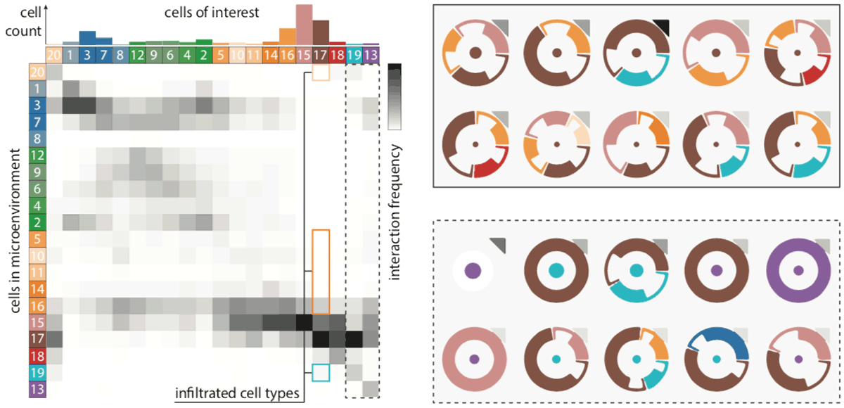
Tissue functionality is determined by the characteristics of tissue-resident cells and their interactions within their microenvironment. Imaging Mass Cytometry offers the opportunity to distinguish cell types with high precision and link them to their spatial location in intact tissues at sub-cellular resolution. This technology produces large amounts of spatially-resolved high-dimensional data, which constitutes a serious challenge for the data analysis. We present an interactive visual analysis workflow for the end-to-end analysis of Imaging Mass Cytometry data that was developed in close collaboration with domain expert partners. We implemented the presented workflow in an interactive visual analysis tool; ImaCytE. Our workflow is designed to allow the user to discriminate cell types according to their protein expression profiles and analyze their cellular microenvironments, aiding in the formulation or verification of hypotheses on tissue architecture and function. Finally, we show the effectiveness of our workflow and ImaCytE through a case study performed by a collaborating specialist.
Resources
Citation
BibTeX
@article{ bib:2019_imacyte,
    author = {Antonios Somarakis and Vincent van Unen and Frits Koning and Boudewijn Lelieveldt and Thomas H{\"o}llt},
    title = { ImaCytE: Visual Exploration of Cellular Microenvironments for Imaging Mass Cytometry Data },
    journal = { IEEE Transactions on Visualization and Computer Graphics },
    volume = { 27 },
    number = { 1 },
    pages = { 98 -- 110 },
    year = { 2021 },
    doi = { 10.1109/TVCG.2019.2931299 },
}